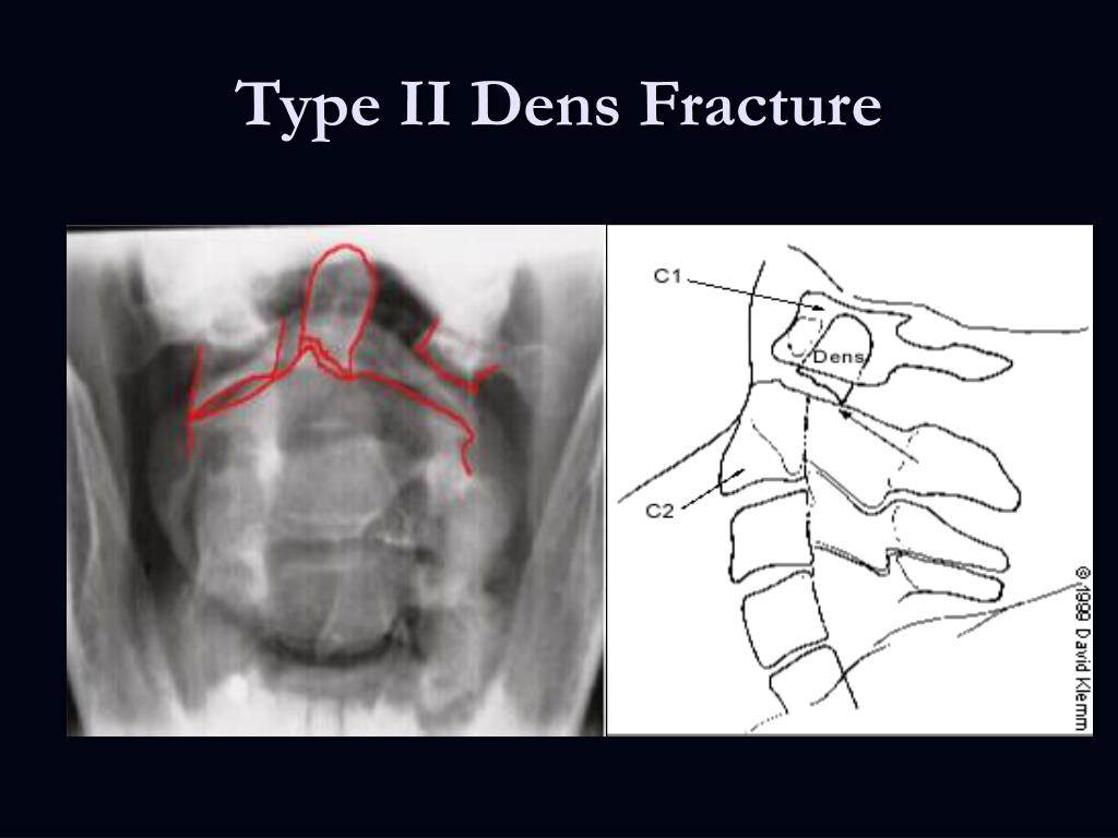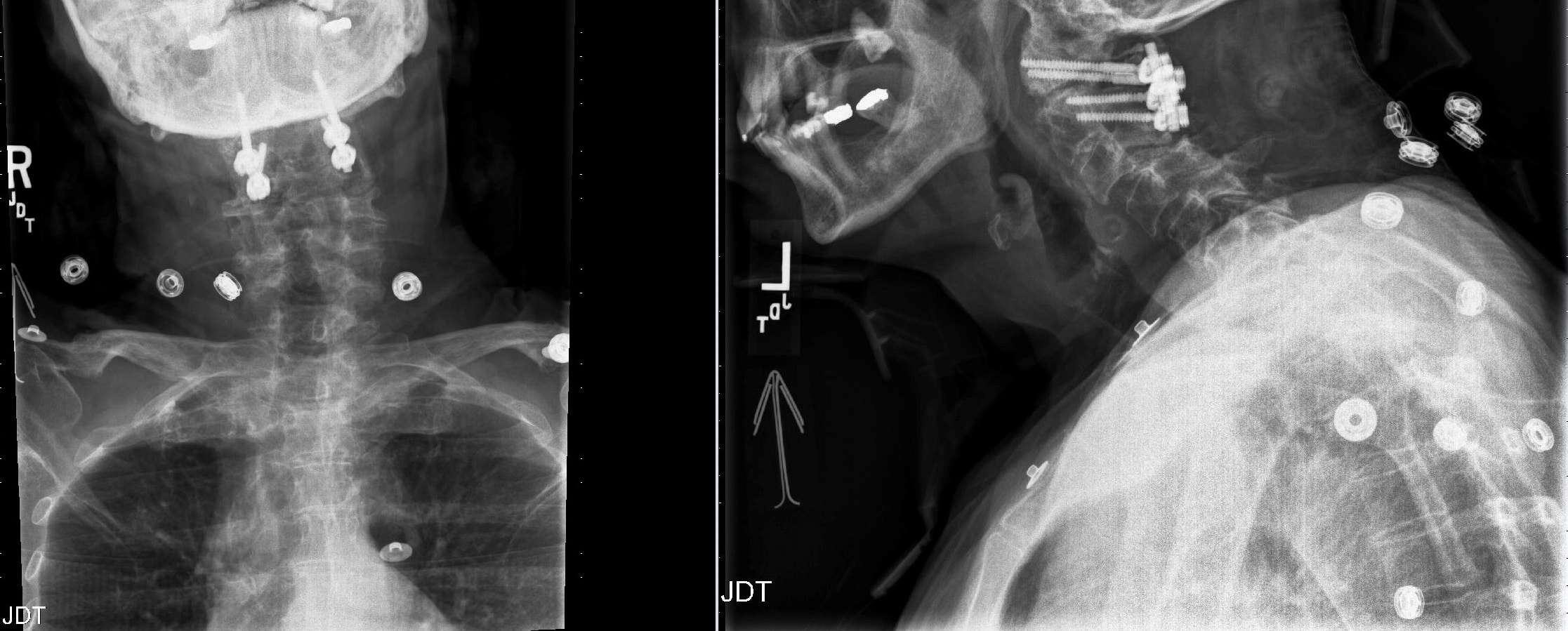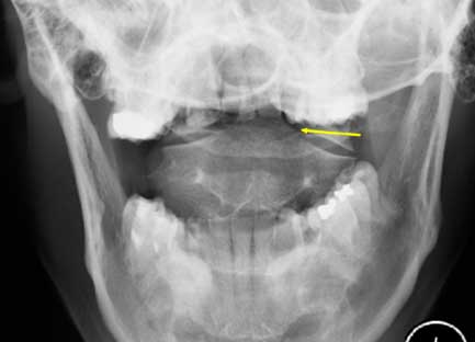
Ĭomputerized tomography (CT) spine - provides the best resolution of the bony elements allowing for the identification and characterization of an odontoid fracture. Initial stability on upright radiographs is associated with stability on follow-up. In addition, flexion-extension radiographs should be obtained in the setting of suspected occipitocervical instability (useful in type I odontoid fractures or the setting of os odontoideum). Although radiographs yield lower sensitivity and specificity rates when compared to computed tomogram (CT) scans, experienced clinicians and practitioners can still appreciate suspected injury without CT utilization. X-rays of the cervical spine constitute lateral, anterior-posterior (AP), and open-mouth views. There are fewer spinal cord injuries in odontoid fractures due to the relatively large cross-sectional diameter of the spinal canal at the level of the odontoid process compared to the diameter of the spinal cord. Less commonly, the patient may have myelopathic spinal cord injuries such as paresthesias in the arms and/or legs, weakness of the arms and/or legs, or other neurologic dysfunctions. They can also have dysphagia due to a retropharyngeal hematoma or associated parapharyngeal swelling.

On physical exam, patients may note cervical neck pain, which is worse with motion. However, older individuals can also sustain an odontoid fracture from recent injuries similar to those of younger people. Older patients tend to have less resilient bones and can sustain an odontoid fracture after minor trauma, including falling from ground level or running into a door or cabinet. Younger patients with an odontoid fracture typically have identifiable recent trauma (motor vehicle accident, sports-related impact, diving accident, fall from a height or downstairs). In a study comprising more than 30,000 patients, the average age of the cohorts was 77, and 54% were females.

Road traffic accidents are accountable for the majority of cases. Type III odontoid fractures make up most of the remaining odontoid fractures. Type II is the most common of the types of odontoid fractures and accounts for over 50% of all odontoid fractures. Hyperextension injury causes the skull and C1 arch to cause traction at the odontoid process, while the hyperflexion injury causes the transverse ligament to limit the posterior movement of dens. This results from high-impact forces in young cohorts but with trivial injuries among elders. Odontoid fractures result from an interaction between the load magnitude and bone quality. This shows bimodal distribution with peaks among early adults and the elderly population. The axis (C2) is the most common vertebra to be involved in cervical spine injuries, and odontoid fractures account for 50% of all C2 fractures. Odontoid fractures account for 20% of cervical spine fractures in the adult population and are the most common fracture subtype in geriatric patients (≥ 65 years). If the cervical spine is excessively flexed, then the transverse ligament can transmit the excessive anterior forces to the odontoid process and cause an odontoid fracture. The transverse ligament runs dorsal to (behind) the odontoid process and attaches to the lateral mass of C1 on either side. The odontoid fracture can also occur with hyperflexion of the cervical spine. The most common mechanism of injury is a hyperextension of the cervical spine, pushing the head and C1 vertebrae backward. If the energy mechanism and resulting force are high enough (or the patient's bone density is compromised secondary to osteopenia/osteoporosis), the odontoid will fracture with varying displacement and degrees of comminution.

In the elderly population, the trauma can occur after lower energy impacts such as falls from a standing position. In younger patients, they are typically the result of high-energy trauma, which occurs as a result of a motor vehicle or diving accident. Odontoid fractures occur as a result of trauma to the cervical spine.


 0 kommentar(er)
